Diagnostics & Patient Monitoring
Clinical Patient Monitoring in Primary Care: Accurate Blood Pressure, ECG & Pulse Oximetry
21
Jun
Jun
Accurate vital signs measurement is the foundation of patient triage and clinical decision-making. Minor errors in cuff selection, sensor positioning, or skin preparation can produce significant diagnostic artefacts, leading to inappropriate clinical interventions or delayed care.
1. Non-Invasive Blood Pressure (NIBP) Precision Protocols
To avoid false hypertensive or hypotensive readings during patient intake:
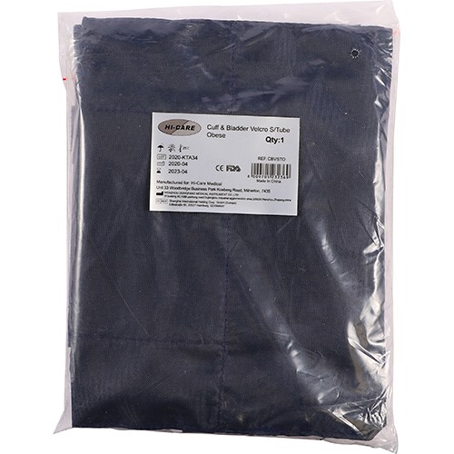
- Bladder Dimension Sizing: The inflatable bladder must encircle at least 80% of the patient’s upper arm circumference, with a width equal to 40% of the upper arm length. A cuff that is too small falsely elevates systolic and diastolic readings by 10–15 mmHg.
- Limb Level: Ensure the cuff is positioned at mid-sternum level (heart level). Every 5 cm the arm is positioned below the heart adds ~4 mmHg of hydrostatic pressure artefact.
- Rest Period: Allow the patient to sit quietly for at least 5 minutes prior to recording baseline measurements. Avoid talking during deflation.
2. Pulse Oximetry (SpO₂) Principles and Limitations
Pulse oximeters calculate arterial oxygen saturation by measuring differential absorption of red (660 nm) and infrared (940 nm) light across pulsatile capillary beds. Clinicians should recognize key sources of reading distortion:
| Clinical Factor | Effect on SpO₂ Reading | Corrective Action |
|---|---|---|
| Poor Peripheral Perfusion / Hypothermia | Loss of pulse waveform signal, erratic values | Warm digit, utilize earlobe or forehead sensor probe |
| Dark Nail Polish / Artificial Acrylic Nails | Falsely depresses SpO₂ (absorbs 660 nm light) | Position sensor horizontally across digit or remove polish |
| Carbon Monoxide Poisoning (Carboxyhaemoglobin) | Falsely normal / high SpO₂ (~98–100%) | Perform Arterial Blood Gas (ABG) co-oximetry analysis |
| Ambient Light Interference (Surgical Lamps) | Optical sensor saturation and fluctuating readings | Cover sensor site with an opaque barrier or cloth |
3. Electrocardiogram (ECG) Skin Preparation and Lead Placement
To eliminate 50 Hz mains baseline interference and muscle tremor artefacts during 3-lead and 12-lead recordings:
- Cleanse electrode sites with an alcohol swab and allow to dry thoroughly.
- Gently abrade the epidermis to lower skin contact impedance.
- Ensure electrode gel is moist; never use dried or expired disposable electrodes.
- Apply limb leads over bony prominences rather than active muscle bellies.